Researchers at Revel Pharmaceuticals, working with collaborators at Calico Life Sciences and the University of Colorado Anschutz Medical Campus, recently reported “Reversal of Protein Chemical Aging by Enzymatic Deglycation” in Nature Communications. Their study introduced CrGO-897, or CMLase: an engineered glycine oxidase that removes Nε-carboxymethyl-lysine (CML) from peptides, proteins, and aged human tissue samples. CML is an advanced glycation end product that accumulates on long-lived extracellular matrix proteins.
We independently asked a narrower computational question: what might a reaction-compatible complex between CrGO-897, FAD, and peptide-bound CML look like?
Why ECM damage matters
The extracellular matrix is more than structural scaffolding. It provides biochemical and mechanical signals that influence how cells grow, migrate, differentiate, and respond to injury. Changes to the matrix can therefore alter both tissue mechanics and cell behavior.
Many ECM proteins, including collagen and elastin, turn over slowly and can remain in the body for years or decades. That longevity makes them especially vulnerable to cumulative chemical damage. Advanced glycation end products form through nonenzymatic reactions and build up with age, particularly in tissues such as skin, arteries, kidneys, cartilage, and the eye.
CML is one of the most abundant of these modifications. It converts a positively charged lysine side chain into an anionic adduct and can alter local protein interactions. CML-modified proteins can also activate the receptor for advanced glycation end products (RAGE), linking accumulated matrix damage to inflammation and oxidative stress. AGE accumulation is associated with aging as well as diabetes, vascular disease, kidney disease, fibrosis, and loss of tissue elasticity.
Why repairing the ECM is difficult
Reversing an isolated chemical modification is only part of the problem. A useful repair enzyme must work in a dense, heterogeneous environment where every modified lysine has a different local structure and degree of solvent exposure.
- Access: modified residues may be buried inside a folded protein or shielded within a dense, cross-linked matrix.
- Specificity: the enzyme must recognize CML without damaging unmodified amino acids or related modifications.
- Delivery: activity on purified proteins or thin tissue sections does not establish penetration into an intact, living organ.
- Functional recovery: removing CML chemically does not yet prove that tissue mechanics or RAGE signaling will be restored.
- Biological breadth: CML is only one member of a chemically diverse family of age-related modifications.
These challenges make substrate recognition important. Understanding how CrGO-897 holds peptide-bound CML near FAD can help connect the study’s experimental activity measurements to an atomic-level structural hypothesis.
Independent analysis
This work is based on the published enzyme design and experimental foundation. The original authors were not involved in this computational follow-up.
Computational methods
Sequence reconstruction and structural model
We started from the 301-residue wild-type Calidithermus roseus glycine oxidase sequence (UniProt A0A399F051). The published mutation history was applied cumulatively, including the reported two-residue deletion, to reconstruct the 299-residue CrGO-897 sequence. Every edited position was checked against the wild-type sequence before the final model was submitted to AlphaFold Server with templates enabled.
No experimental structure of CrGO-897 was reported. We therefore aligned the predicted model to the FAD/N-acetylglycine-bound Bacillus subtilis glycine oxidase structure, PDB 1NG3, and transferred the crystallographic FAD coordinates into the CrGO-897 model. The acetylglycine carbon in 1NG3 lies 3.45 Å from FAD N5 and served as a structural reference—not as proof of the CrGO-897 reaction geometry.
Protein, FAD, and peptide preparation
The protein was prepared at pH 8 using PDB2PQR with the AMBER force field and PROPKA-assigned protonation states. FAD bond orders were restored from the PDB chemical-component definition and prepared in its pH-8, net −2 charge state.
The ligand matched the paper’s AA[CML]AA model substrate. We built it as an all-L peptide with ACE and NME terminal caps to represent an internal protein segment without artificial charged termini. For this trajectory, the CML secondary amine was protonated and its carboxylate was deprotonated, giving the capped ligand a net charge of zero.
Both the nonstandard CML peptide and FAD were parameterized with
AmberTools. Antechamber assigned GAFF2 atom types
and AM1-BCC partial charges, while Parmchk2 generated missing bonded
terms. The protein used the AMBER ff14SB force field. This mixed
ff14SB/GAFF2 parameter set was assembled and checked with
tleap.
Holo docking and geometry-based pose selection
FAD was kept as a rigid part of the receptor during docking so that the peptide could not occupy the empty flavin cavity. We used AutoDock Vina 1.1.2 with a 30 × 30 × 30 Å search box centered on the transferred 1NG3 substrate site, an exhaustiveness of 64, a fixed random seed of 42, and 20 output poses.
In addition to the Vina score, each pose was evaluated using two mechanistic geometric criteria: the distance from the CML carboxymethyl carbon to FAD N5, and the displacement of that carbon from the acetylglycine reference position in 1NG3. This second step is what identified pose 19.
The best score missed the chemistry
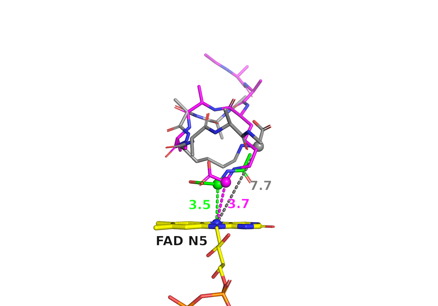
The highest-ranked pose scored −7.0 kcal/mol, but placed the CML carboxymethyl carbon 7.70 Å from FAD N5. The peptide fit the pocket, yet its proposed hydride-donor carbon was poorly positioned for the expected flavin chemistry.
Pose 19 scored only 0.6 kcal/mol worse, within the practical uncertainty of a docking score, but placed the same carbon 3.69 Å from FAD N5. It was also within 0.73 Å of the substrate-carbon position observed in the homologous 1NG3 crystal structure.
Binding score alone is not a proxy for a reaction-compatible enzyme–substrate geometry.
Testing the pose with molecular dynamics
Pose 19 was converted into an Amber topology with
tleap. The complex used ff14SB for CrGO-897, GAFF2 for
FAD and the capped CML peptide, and TIP3P water in a truncated
octahedral box extending 10 Å from the solute. Sodium and chloride
ions were added to neutralize the approximately 41,800-atom system.
We confirmed after system construction that the starting
carboxymethyl-carbon–N5 distance remained 3.69 Å.
Molecular dynamics was run with OpenMM. The system was energy-minimized, followed by 200 ps of restrained NPT equilibration with heavy-atom restraints on the solute and a brief unrestrained settling stage. Production consisted of one 2 ns unrestrained NPT trajectory at 300 K, using a 2 fs timestep, particle-mesh Ewald electrostatics, and coordinates saved every 10 ps. The calculation ran through OpenMM’s OpenCL platform.
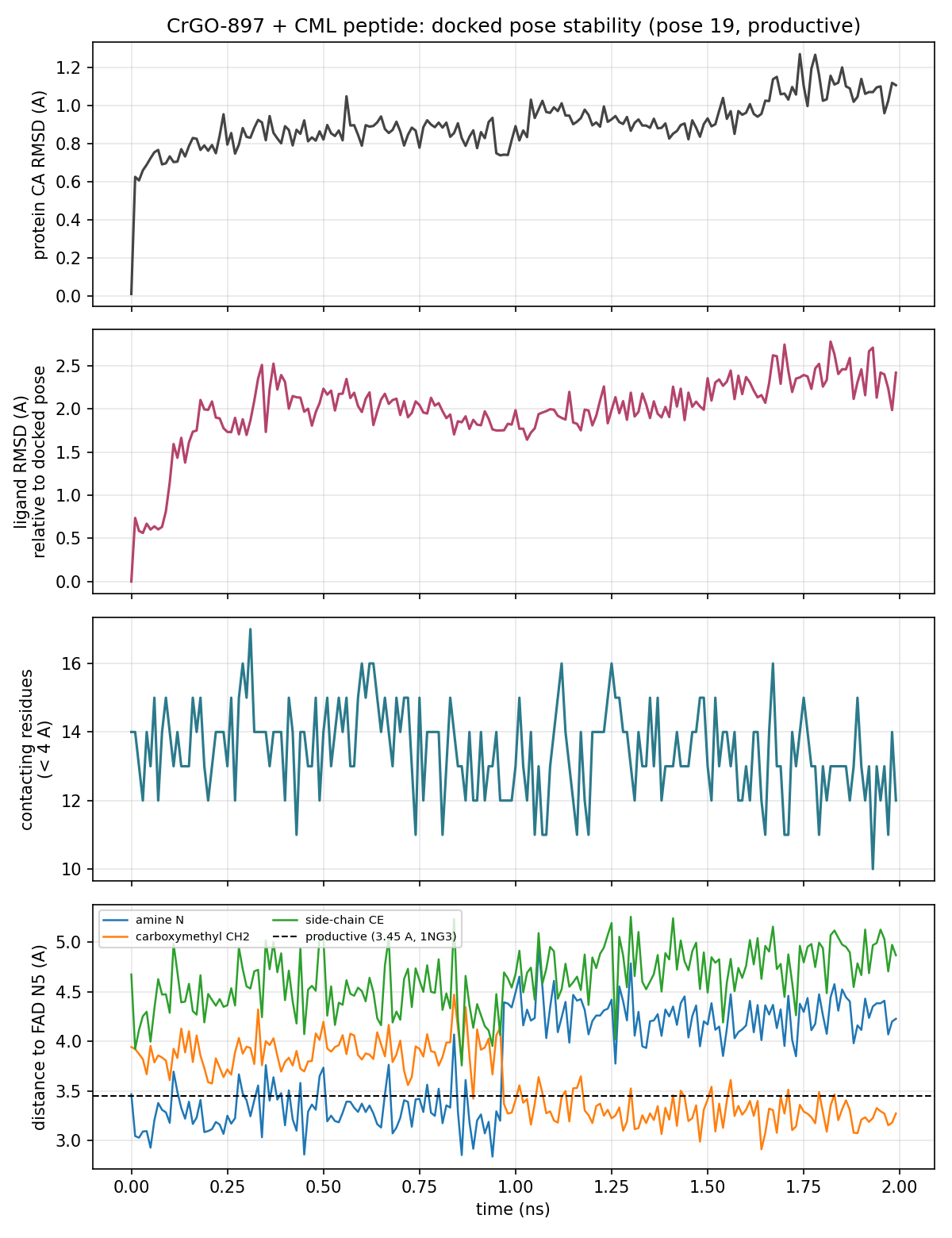
Trajectory analysis measured protein Cα RMSD, ligand and FAD heavy-atom RMSD, protein–ligand contacts within 4 Å, and the distances from three candidate CML atoms to FAD N5. The carboxymethyl carbon was the reaction-relevant coordinate for the proposed deglycation chemistry.
Results from one 2 ns trajectory
- Mean CML carboxymethyl carbon–FAD N5 distance: 3.58 Å
- Second-nanosecond distance: 3.29 ± 0.14 Å
- Mean protein Cα RMSD: 0.90 Å
- Mean ligand heavy-atom RMSD: 1.99 Å
- Mean FAD heavy-atom RMSD: 0.72 Å
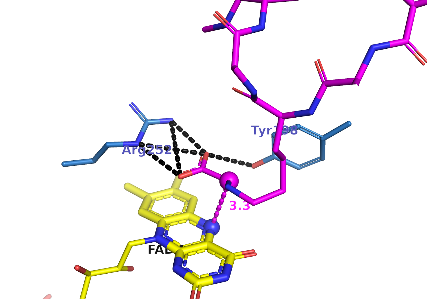
The paper identified several pocket residues that improved CML activity through directed evolution. Our trajectory analysis adds a structural hypothesis by identifying Arg252 and Tyr198 as persistent contacts that may help orient the CML carboxylate near FAD.
What this result does—and does not—show
This simulation supports a structural hypothesis, not an experimental claim of catalysis. Classical MD cannot model hydride transfer or bond breaking. The analysis also used a predicted CrGO-897 structure, a transferred cofactor geometry, one short trajectory, and a protonated CML amine model.
Longer replicate simulations, neutral-amine parameterization, and QM/MM calculations would be appropriate next steps. Still, this study demonstrates why mechanistically relevant distances and orientations should be evaluated alongside docking scores—especially when modeling enzymes rather than binders.
Working on ECM Aging or Repair?
SimAtomic works on molecular simulations of the extracellular matrix, including AGE-modified residues, collagen crosslinks, enzyme–matrix recognition, and how chemical damage changes protein structure and dynamics. If you are developing an ECM repair strategy or investigating age-related matrix damage at atomic resolution, we would be glad to discuss your project.
Discuss an ECM ProjectReferences
- Trabosh N, Smith J, Hsu MYH, et al. (2026). “Reversal of protein chemical aging by enzymatic deglycation.” Nature Communications 17, 5926. DOI
- Settembre EC, Dorrestein PC, Park J, et al. (2003). “Structural and mechanistic studies on ThiO, a glycine oxidase essential for thiamin biosynthesis in Bacillus subtilis.” Biochemistry 42, 2971–2981. DOI